Skip to content
  • Stanford Medicine
  • •
  • Otolaryngology — Head & Neck Surgery
Skull Base Surgery Atlas Logo
  • Home
  • About this Atlas
Illustrations of Skull Base Surgery and NeurotologyScott Stocker2023-01-23T09:35:11-08:00
An axial view of the skull through the level of the of the internal auditory canal and cerebellopontine angle.

Surgical and Tumor Anatomy

Axially oriented schematic view of a translabyrinthine posterior fossa craniotomy used in the exposure of a medium-sized acoustic neuroma. Note that bone has been removed 1 to 2 cm behind the sigmoid sinus which is retracted posteriorly. The amount of cerebellar retraction is considerably less than that required with the retrosigmoid approach. Note that the semicircular canals have been removed en route to exposing the internal auditory canal.

Retro- labyrinthine, Trans- labyrinthine, Transcochlear Approaches

In the classical suboccipital approach bone is removed from the midline laterally until the first air cells are encountered. This often creates an opening located well posterior to the sigmoid sinus. When a suboccipital approach is used to expose the CPA, a relatively large amount of cerebellar retraction is necessary. With the retrosigmoid approach, where the opening created immediately adjacent to the sigmoid and transverse sinuses, much less cerebellar displacement is required. (SO, suboccipital; RS, retrosigmoid.)

Retrosigmoid Approach

Completed exposure of a left translabyrinthine posterior fossa craniotomy before dural opening. (PF, posterior fossa; MF, middle fossa; IAC, internal auditory canal.)

Microsurgery of Acoustic Neuroma

(A) A schematic coronal view through the temporal bone illustrating the relationship between the undersurface of the temporal lobe and the roof of the petrous pyramid. An intracanalicular acoustic neuroma is present. Note that this often-used style of illustrating the inner ear in the coronal plane, which makes both the semicircular canals and cochlea visible through a single coronal slice, is inaccurate. The cochlea actually lies just beneath the floor of the middle cranial fossa.

Middle Fossa Approach

The classical method of exposing both the posterior and middle cranial fossae is by creating two separate openings – retrosigmoid and subtemporal. This disconnected approach, with the surgical exposures separated by the transverse sinus superficially and the tentorium in the depth, necessitates a much greater degree of brain retraction than contemporary approaches to tumors involving both fossae.

Combined Craniotomies: Posterior & Middle Fossae

Completed exposure of a meningioma involving both the middle and posterior fossa by traversing Meckel’s cave via the combined retrolabyrinthine-middle fossa approach. The tumor spans from the entry zone of the audiovestibular and facial nerves (7,8) to the cavernous sinus (CS). Note the trochlear nerve on the tumor surface (4). The distil of the branches of the trigeminal nerve can be seen (V2, V3). The posterior retractor is on the cerebellum (Cb). The superior retractor is on the temporal lobe (TL). The curled dural flap containing the endolymphatic sac (ES) overlies the petrous portion of the temporal bone containing the semicircular canals. All three segments of the brainstem are visible: medulla (M), pons (P), midbrain (MB). The edges of the divided tentorium (T) can be seen anteriorly and posteriorly. (for details of the microsurgical exposure, see “Combined Craniotomies of the Posterior and Middle Fossae).

Meningioma via Combined Approach

Anatomical overview of the jugular foramen region and its neurovascular relationships. (9, glossopharyngeal nerve; 10, vagus nerve; 11, spinal accessory nerve; 13, hypoglossal nerve.)

Jugular Foramen Approach

Meningiomas involving the foramen magnum region may arise either from the anterior or posterior aspect of this osseous ring. They are often craniovertebral in that they extend from the posterior fossa into the upper spinal column. Posteriorly placed lesions can be removed in a relatively straight forward manner by opening the posterior aspect of the ring. Removal of anteriorly situated lesions, which often arise from the lower 1/3 of the clivus, are much more challenging. The ventral position of these lesions lies against the anterior face of the lower medulla and upper spinal cord and often involve the vertebrobasilar system. A lateral approach necessitates removal of a portion of the occipital condyle. Hence the alternative term for this approach: the transcondylar approach. Anterior approaches, which traverse the pharynx, are generally not considered suitable for intradural lesions in this location. (A, anterior; P, posterior; C, clivus, C1, first cervical vertebra; C2, second cervical vertebra; FM, foramen magnum; O, odontoid.)

Foramen Magnum Approach

(A) The orifice of Meckel’scave (Cavum trigeminale) as seen from the posterior fossa. Distal to the semilunar ganglion the nerve divides into its three main branches as depicted here. The dural surfaces related to Meckel’scave (violet) include: the tentorium (green), the posterior petrous face (yellow), the clivus (orange), and the lateral wall of the cavernous sinus (blue).

Meckel’s Cave Approaches

View of the cranial base as seen from above. The clivus is a portion of the occipital bone which extends from the anterior margin of foramen magnum to the dorsum sellae.

Approaches to the Clivus

Figure 12-1 Position and incision (dashed line). The patient is placed supine and the ipsilateral shoulder is elevated to permit turning of the head into the lateral position. A Mayfield headholder is placed and the head is rotated 20 degrees ipsilaterally and tilted down 20 degrees to bring the pterion uppermost. The incision is designed to expose the temporal muscle while preserving the posterior branch of the superficial temporal artery and the temporal and zygomatic branches of the facial nerve. It begins in the pretragal crease just below the root of the zygoma and passes superiorly across the superior temporal line behind the coronal suture before arcing anteriorly to the midline hairline. A receding hairline may require a full bicoronal incision.

Orbitozygomatic approaches

Endoscopic endonasal approach to the central skull base. The divergent light of an endoscope placed in one nostril illuminates a broad area of the anterior and central skull base. Straight and angled endoscopes permit access to the frontal sinus, cribriform plate, ethmoid roof, sphenoid sinus, planum sphenoidale, sella, pituitary gland, suprasellar region, cavernous sinuses, dorsum sella, and midclival regions. This facilitates repair of encephaloceles and removal of tumors such as esthesioneuroblastomas, meningiomas, pituitary tumors, and clival chordomas.

Use of Endoscopes in Skull Base Surgery

Schematic illustration of the major arterial branches in the cerebellopontine angle. (IAA, internal auditory artery; SCA, superior cerebellar artery; AICA, anterior inferior cerebellar artery; PICA, posterior inferior cerebellar artery.)

Vascular Considerations in Lateral Skull Base Surgery

Fractures through the petrous pyramid may result from either posterior or lateral blows to the head. The skull base fractures more easily when struck from the side, on the thin temporal squamosa, than posteriorly on the thick occipital bone. Fractures resulting from temporal blows tend to propagate medially along a course parallel to the long axis of the petrous pyramid and thus are termed longitudinal. Strong occiptal blows characteristically produce a fracture which disrupts the foramen magnum ring and then propagates anteriorly across the petrous pyramid art right angles to its long axis. These are termed transverse fractures.

Fractures of the Cranial Base

Encephaloceles of the temporal bone most commonly occur through the floor of the middle cranial fossa into the epitympanum or mastoid. Most frequently, they are traumatic in origin, arising either due to cranial base fracture or after iatrogenic injury sustained during mastoid surgery. Spontaneous meningoceles and encephaloceles also occur, presumably due to prominent arachnoid granulations that penetrate the eggshell-thin tegmen mastoideum or tympani. Chronically increased intracranial pressure may play a role in such cases.

Encephalocoele

Management of cerebrospinal fluid (CSF) otorhinorrhea illustrated by leakage occurring after a translabyrinthine approach to acoustic neuroma. In this case, CSF traverses the fat graft in the petrous defect to pass through the aditus to the middle ear and then via the eustachian tube to the nasopharynx (arrow).

Cerebrospinal Fluid Leak

Temporal bone resection for malignancy encompasses three related procedures of progressively increasing depth: sleeve resection of the external auditory canal (solid line), lateral temporal bone resection (dotted line), total temporal bone resection (dashed line). The illustration depicts these procedures in the coronal view. Most of these resections are performed for squamous cell carcinoma arising from the external auditory canal. It is generally acknowledged that sleeve resection is insufficient therapy for malignant disease. In the lateral temporal bone resection, the ear canal is removed en bloc with the tympanic membrane and lateral ossicles. A parotidectomy and/or neck dissection often supplements the temporal bone specimen. In total temporal bone resection, creation of an en bloc specimen is difficult and probably unnecessary. It requires an extensive dissection of the intrapetrous carotid artery, a measure usually of little benefit in deeply invasive squamous cell carcinoma. Most contemporary otologists perform the so-called total temporal bone resection by first carrying out a lateral temporal bone resection and then removing the medial petrous bone piecemeal with a drill. This procedure is indicated for deep extension which penetrates beyond the medial wall of the middle ear and/or mastoid.

Temporal Bone Resection

The petrous apex is the medial portion of the petrous bone that lies between the inner ear and the clivus. Petrous apicectomy is the term applied to a procedure which reaches the apical portion of the petrous bone by skirting around the inner ear. It is inherently a drainage procedure which creates only a relatively small entry into the apical region. Thus, the commonly used term petrous apicectomy is a misnomer when used in this context. Petrous apicotomy would actually be a more accurate description for the procedure. Petrous apicectomy is primarily indicated for drainage of cholesterol granuloma and purulent infections. Fundamentally, there are two routes used to reach the petrous apex: those which pass near the labyrinth and those which skirt the cochlea. In recent years, the hypotympanic - subcochlear route depicted here has become the most popular. The bone removed during this procedure is depicted in this schematic coronal illustration as the color green. Note the relationship of the apical cholesterol granuloma to the fifth and sixth nerves. This explains the frequent occurrence of deep ear and retro-orbital pain as well as diplopia in these lesions. (JV, jugular vein; CA, carotid artery; CG, cholesterol granuloma; 5, trigeminal nerve; 6, abducens nerve.)

Approaches to the Petrous Apex

The goal of ear canal closure during skull base surgery is to create a sealed cavity into which an adipose tissue graft or muscle flap can be placed. Prior to obliterating the surgical defect, all skin and as much mucous membrane as possible must be removed from the defect. When the dura has been violated, it is essential that the closure be watertight to avoid cerebrospinal fluid leakage. This requires a multiple layer closure and meticulous technique. It is usually done in the early stages of the procedure, although it can be accomplished at any convenient stage. The ear canal is initially transected at the level of the bony cartilage junction (dashed line). For the technique of meatal closure employed in temporal bone resection for carcinoma of the ear canal, please see Chapter 5.

Ear Canal Closure in Skull Base Surgery

Schematic overview of the anatomy of the facial nerve course from the brainstem (A) to its terminal branches at the facial muscles (B).

Facial Nerve Surgery

The facial nerve seen in its normal anatomic position within the temporal bone seen after completion of a canal wall down mastoidectomy (subtotal petrosectomy). Note than the descending portion of the nerve in the mastoid overlies the jugular foramen. (1G, first genu; 2G, second genu; SMF, stylomastoid foramen; JB, jugular bulb; CA, carotid artery.)

Facial Nerve Rerouting during Skull Base Surgery

Hypoglossal - facial anastomosis is performed through a pre-auricular incision which is carried into the upper neck approximately 2 cm beneath the mandible (dashed line). (7, facial nerve; 12, hypoglossal nerve; HB, hyoid bone.)

Reanimation of the Paralyzed Face

Selective vestibular neurectomy may be performed either proximally, near the brainstem root entry zone, or distally in the internal auditory canal. Neurectomy in the posterior fossa may be performed through either a retrosigmoid exposure (as depicted here) or via the retrolabyrinthine approach (see section 2.3 in Chapter 2). Neurectomy in the internal auditory canal may be accomplished through a middle fossa exposure or, when hearing is absent, via a translabyrinthine approach (see section 2.1 in Chapter 2).

Vestibular Surgery

The placement of the cochlear implant receiver/ stimulator (dotted line) should be planned to allow sufficient room between the pinna and the headpiece to enable the comfortable use of a behind-the-ear processor. Approximately 3 to 4 cm should be left between the ear canal and the anterior edge of the device. The device is angled up at about a 45-degree angle superior to the linea temporalis A modified post-auricular incision is used most commonly. Instead of continuing superior to the pinna, the incision can extend into the supra-auricular hairline to facilitate later drilling of the bony well for the receiver/ stimulator case. The length of this extension depends on surgeon preference and the benefit that additional exposure provides when securing the receiver/ stimulator to bone. Sufficient space (at least 1.5 cm) between the incision and the implant should be allowed to minimize the chance of device extrusion through the incision. Minimal shaving of hair is required if drapes are secured sufficiently to adjacent skin.

Cochlear Implantation

There are numerous reconstructive options for the repair of small defects in the region of the temporal bone. These include local flaps which borrow skin from either the scalp, upper neck, or face. The simplest solution for a limited postauricular defect is a scalp rotation flap as shown here.

Reconstruction of the Cranial Base

The cranial nerve monitoring montage utilized during surgery of the cerebellopontine angle includes electrodes placed in the muscles innervated by each motor nerve in the region (cranial nerves 5, 7, and 11). To monitor the facial nerve, electrode pairs are inserted into both the orbicularis oris and oculi muscles. The motor division of the trigeminal is monitored through an electrode pair in the temporalis muscle.

Cranial Nerve Monitoring

From ATLAS OF SKULL BASE SURGERY & NEUROTOLOGY. Thieme. ©2009

All images are copyright by RK Jackler. Permission granted for non-profit educational use of images, with attribution to their source.

Created by: Robert Jackler (surgeon) and Christine Gralapp (artist).

With contributions by:
Nikolas Blevins, Griffith Harsh, Michael Kaplan, Lawrence Pitts, Charles Yingling, and Corey Mass.

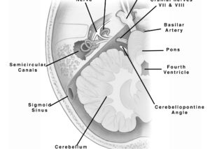
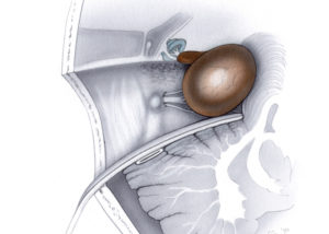
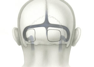
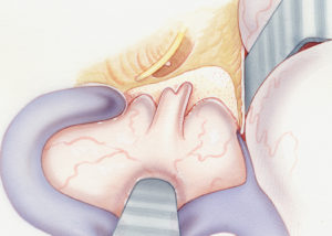
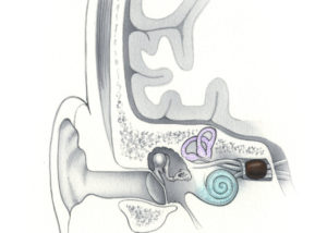
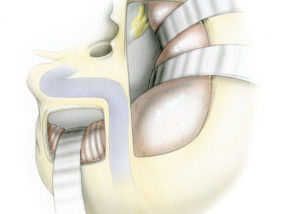
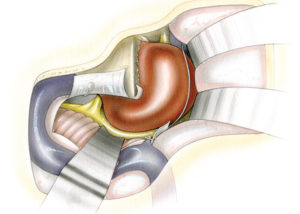
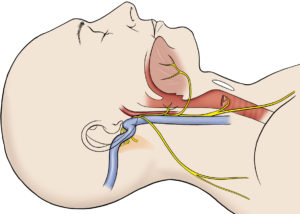
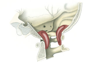
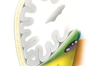
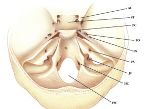
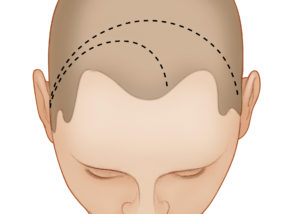
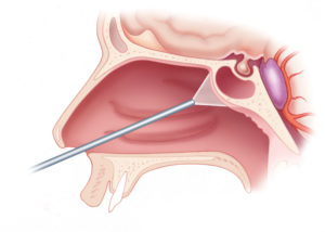
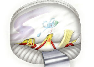
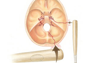
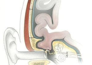
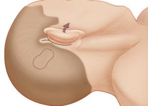
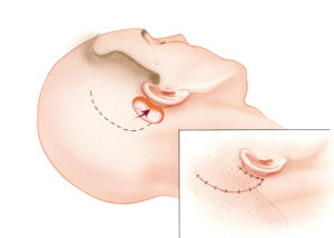
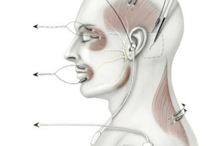
For legend references, please see the print book.

  • Illustrations of Skull Base Surgery and Neurotology
  • Surgical and Tumor Anatomy
    • Surgical Anatomy
    • Anatomy of Tumors in Skull Base Surgery and Neurotology
  • Retro- labyrinthine, Trans- labyrinthine, Transcochlear Approaches
    • Translabyrinthine approach
    • Transcochlear approach
    • Retrolabyrinthyine approach
  • Retrosigmoid Approach
  • Microsurgery of Acoustic Neuroma
  • Middle Fossa Approach
    • Middle Fossa Approach to the Internal Auditory Canal
    • Extended Middle Fossa Approach to the Cerebellopontine Angle
  • Combined Craniotomies: Posterior & Middle Fossae
    • Combined transpetrosal – middle fossa approaches
    • Middle fossa transpetrous apex approach to the anterosuperior CPA
  • Meningioma via Combined Approach
  • Jugular Foramen Approach
    • Jugular Foramen Approaches
    • Transjugular Craniotomy
    • Retrosigmoid Approach to Jugular Foramen
    • Infratemporal Fossa Approaches
  • Foramen Magnum Approach
  • Meckel’s Cave Approaches
    • Posterior Fossa Approach
    • Middle Fossa Approach
    • Combined Middle and Posterior Fossa Approach
  • Approaches to the Clivus
    • Anterior Approaches to Clivus
    • Lateral Approaches to Clivus
  • Orbitozygomatic approaches
  • Use of Endoscopes in Skull Base Surgery
  • Vascular Considerations in Lateral Skull Base Surgery
  • Fractures of the Cranial Base
  • Encephalocoele
  • Cerebrospinal Fluid Leak
  • Temporal Bone Resection
    • Standard Procedure
    • Extended Temporal Bone Resection
  • Approaches to the Petrous Apex
    • Petrous Apicotomy: Infracochlear and Infralabyrinthine Approaches
    • Petrous Apicectomy: Middle Fossa Approach
  • Ear Canal Closure in Skull Base Surgery
  • Facial Nerve Surgery
    • Facial Nerve Anatomy
    • Cholesteatoma and the Facial Nerve
    • Facial Nerve Repair
  • Facial Nerve Rerouting during Skull Base Surgery
  • Reanimation of the Paralyzed Face
    • Hypoglossal-Facial Anastomosis
    • Temporalis Muscle Sling
  • Vestibular Surgery
    • Vestibular Neurectomy
    • Endolymphatic Sac Surgery
    • Repair of Superior Semicircular Canal Dehiscence
  • Cochlear Implantation
  • Reconstruction of the Cranial Base
  • Cranial Nerve Monitoring

© Stanford Medicine • Privacy Policy • Terms of Use
Page load link
Go to Top